Microscope
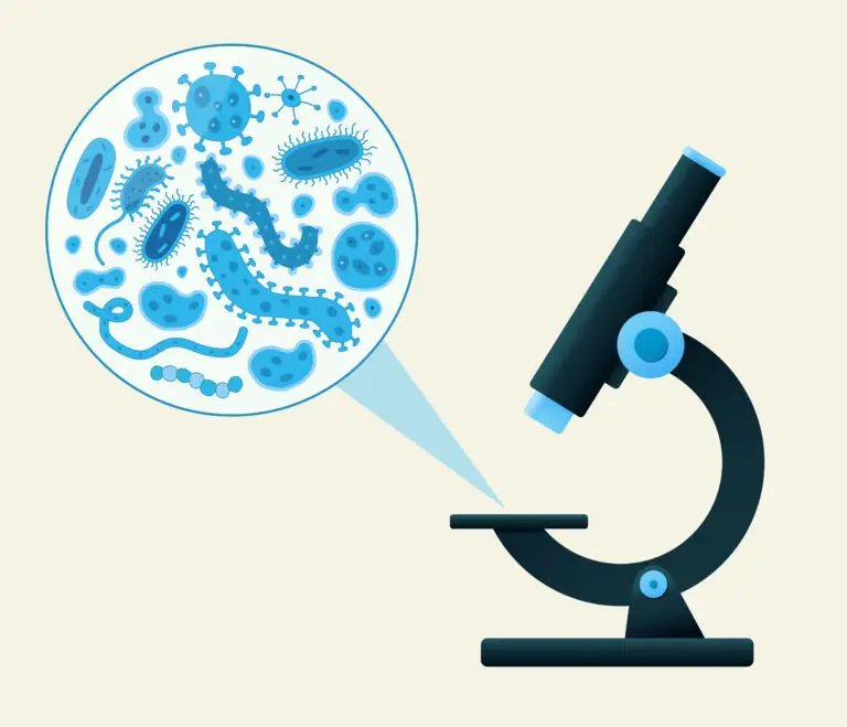
Table of Contents
What is a Microscope?
A microscope is an optical instrument that magnifies small objects or details that are not visible to the naked eye. It allows scientists, researchers, and students to observe and study microscopic structures, such as cells, microorganisms, and other tiny specimens. Microscopes have been crucial tools in advancing our understanding of the biological, medical, and physical sciences.
Components of Microscopes
Objective Lenses
Objective lenses are the primary magnifying components of a microscope. They are positioned near the specimen and contribute to the overall magnification. Microscopes often have multiple objective lenses with different magnification levels.
Eyepiece (Ocular)
The eyepiece, or ocular, is the lens through which the observer looks. It further magnifies the image produced by the objective lens. Microscopes commonly have one or two eyepieces.
Magnification
Microscopes provide total magnification, which is the result of multiplying the magnification of the objective lens by the magnification of the eyepiece. For example, if the objective lens has a magnification of 40x and the eyepiece has a magnification of 10x, the total magnification is 400x.
Illumination System
Microscopes are equipped with an illumination system to provide light for viewing the specimen. This can be achieved through built-in light sources, such as bulbs or LEDs, positioned below the specimen (transmitted light) or above the specimen (reflected light).
Stage and Stage Clips
The stage is the platform where the specimen is placed for observation. Stage clips secure the specimen in place. Some microscopes also have a mechanical stage, which allows precise movement of the specimen.
Focus Mechanism
Microscopes have a focus mechanism that allows users to bring the specimen into sharp focus. This typically involves coarse adjustment for initial focusing and fine adjustment for precise focusing.
Diaphragm
The diaphragm is located beneath the condenser and controls the amount of light reaching the specimen. It can be adjusted to optimize contrast and brightness.
Types of Microscopes
- Compound Microscope: Most commonly used in biology and medicine, it employs multiple lenses to achieve high magnification.
- Stereo Microscope (Dissecting Microscope): Used for three-dimensional viewing of larger specimens, often in dissections or industrial applications.
- Electron Microscope: Uses electron beams instead of light, providing extremely high magnification for detailed imaging of subcellular structures. Includes transmission electron microscopes (TEM) and scanning electron microscopes (SEM).
Related Links
Bacteria
Prokaryote
Quaternary Structure
Virus